Magnetic resonance imaging (MRI) has become an indispensable tool in prenatal diagnostics, offering detailed, non-invasive insights into fetal development. The Vittore Buzzi Children's Hospital in Milan stands out as a leading European center in this field, renowned for its extensive experience and state-of-the-art infrastructure. This article delves into the intricacies of fetal MRI, its applications, benefits, and the specialized services offered at the Buzzi Hospital.
Introduction to Fetal Magnetic Resonance Imaging
Fetal MRI is an advanced imaging technique that utilizes magnetic fields and radio waves to generate high-resolution images of fetal organs and structures. Unlike X-rays or computed tomography (CT) scans, MRI does not employ ionizing radiation, making it a significantly safer option for both the mother and the developing fetus. This safety aspect is paramount in prenatal diagnostics, where the well-being of the fetus is the highest priority.
The initial application of MRI in the fetal context dates back to 1980, primarily for studying the placenta. However, it was not until the 1990s that its potential for examining the fetal brain began to be explored. Significant hurdles, such as prolonged acquisition times and fetal movements causing image artifacts, initially limited its widespread clinical use. The advent of ultra-fast "Single Shot" T2-weighted sequences was a critical advancement, enabling efficient and effective fetal MRI examinations.
The Buzzi Hospital: A Center of Excellence in Fetal MRI
The Vittore Buzzi Children's Hospital has cultivated a unique expertise in fetal MRI, establishing itself as a go-to destination for families and healthcare professionals seeking advanced prenatal imaging. The hospital has performed thousands of fetal MRI examinations, accumulating invaluable experience in diagnosing a broad spectrum of fetal conditions. This expertise, combined with cutting-edge technology and a patient-centered approach, solidifies the Buzzi Hospital's position as a center of excellence.
The Radiology and Pediatric Neuroradiology Unit at the Vittore Buzzi Children's Hospital is the sole autonomous radiology service in Lombardy dedicated exclusively to newborns, children, and pregnant women. The unit offers nearly all types of diagnostic imaging examinations required in pediatric age, excluding certain specialized procedures like scintigraphy, interventional angiography, and arthrography. It functions as a third-level advanced pediatric diagnostic hub.
Key diagnostic activities include high-resolution ultrasound examinations, advanced applications of neonatal and pediatric MRI, and fetal MRI, where the center is recognized as a European leader with over 3,600 examinations performed. The mission is to provide increasingly accurate diagnoses rapidly and with minimal invasiveness, adopting an approach that is as child-friendly as possible. Achieving and maintaining these objectives necessitates continuous professional development, equipment upgrades, environmental humanization, and procedural revisions.
Indications for Fetal MRI
Fetal MRI is not a routine examination; rather, it is employed in specific scenarios where conventional ultrasound, the most common prenatal screening tool, does not provide sufficient information or raises concerns. The primary indications for fetal MRI include:
- Cerebral Anomalies: Fetal MRI is exceptionally useful for evaluating the fetal brain, aiding in the identification of developmental abnormalities, ischemic lesions, hemorrhages, and other neurological conditions.
- Spinal Anomalies: MRI can detect neural tube defects such as spina bifida, offering detailed insights into the severity and extent of the lesion.
- Internal Organ Anomalies: MRI can be utilized to assess abnormalities of the heart, lungs, kidneys, and gastrointestinal tract.
- Placental Anomalies: MRI can assist in identifying placental issues like placenta previa or placenta accreta, which can pose risks to both mother and fetus.
- Multiple Gestations: In cases of twin pregnancies, MRI can be beneficial for evaluating the growth and development of both fetuses and for identifying specific complications associated with multiple births.
- Evaluation of Fetal Masses: When ultrasound reveals a mass within the fetus, MRI can provide detailed information about its nature, size, and location, facilitating treatment planning.
- Hydrocephalus: MRI is a precise method for assessing fetal hydrocephalus, determining the severity of the condition and guiding therapeutic decisions.
- Confirmation or Exclusion of Ultrasound-Suspected Anomalies: In many instances, MRI is used to clarify ambiguous ultrasound findings, leading to a more precise diagnosis.
The Buzzi Hospital's expertise extends to the diagnosis of congenital malformations of the fetal and neonatal body and central nervous system through prenatal MRI and neonatal imaging using ultrasound, CT, contrast-enhanced examinations, and MRI.
Advantages of Fetal MRI
Fetal MRI offers several significant advantages over other prenatal imaging techniques:
- High Resolution: MRI provides high-resolution images, allowing for the visualization of fetal anatomical details with exceptional precision.
- No Ionizing Radiation: Unlike X-rays and CT scans, MRI does not use ionizing radiation, ensuring greater safety for the fetus.
- Multiplanar Assessment: MRI can acquire images in various planes, enabling a comprehensive evaluation of the fetus from multiple angles.
- Soft Tissue Visualization: MRI is particularly effective at visualizing soft tissues, such as the brain, spinal cord, and internal organs.
- Wide Field of View: MRI allows for the visualization of a large area of the fetal body, facilitating the identification of widespread abnormalities.
How a Fetal MRI Examination is Performed
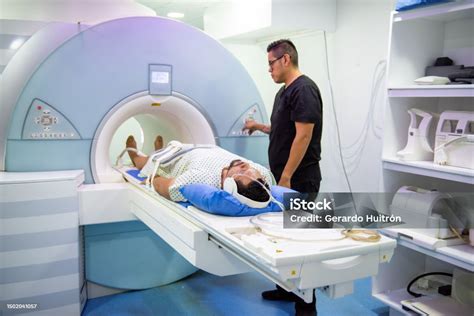
A fetal MRI scan is typically performed between the 18th and 32nd week of gestation. The procedure is painless and non-invasive. The mother lies on a table that is moved into the bore of the MRI scanner. During the examination, it is crucial for the mother to remain as still as possible to minimize image artifacts. In some cases, a mild sedative may be administered to help the mother relax.
The duration of the examination varies depending on the clinical indication and specific diagnostic requirements but generally ranges from 30 to 60 minutes. Throughout the scan, the medical team closely monitors both the mother and the fetus. While MRI is considered safe, a careful assessment of the benefits and risks is necessary for each individual case.
The study conducted at the Vittore Buzzi Hospital analyzed the execution times of 484 fetal brain MRIs performed between January 2015 and December 2017. The analysis aimed to determine if there is a correlation between the total examination time and the gestational age of the patient, with the goal of optimizing examination management, further reducing acquisition times, and enhancing maternal and fetal safety. The findings indicated that as gestational age increases, the duration of the examination tends to decrease. This is attributed to the fact that smaller fetuses exhibit more movement, requiring greater technical skill and experience in setting up scan planes. The "dead time" (the interval between acquired sequences) also tends to decrease with advancing gestational age for this reason.
Magnetic Resonance Imaging (MRI)
Preparation for the Examination
Prior to the MRI scan, the mother will be asked to remove all metallic objects, such as jewelry, watches, and piercings. It is also essential to inform the medical staff about any allergies, pre-existing medical conditions, or implanted devices, such as pacemakers or defibrillators. In some instances, fasting for a few hours before the examination may be required.
Interpretation of Results
The MRI images are interpreted by a radiologist specializing in fetal imaging. The radiologist prepares a detailed report outlining the findings and providing a diagnosis. This report is then forwarded to the referring physician, who will discuss it with the mother and, if necessary, with other specialists, such as a neonatologist or pediatric surgeon.
Clinical and Therapeutic Implications
Accurate prenatal diagnosis through fetal MRI can have significant clinical and therapeutic implications. In some cases, a diagnosis may allow for surgical planning after birth to correct an anomaly. In other situations, it can help parents prepare for the birth of a child with special needs. In certain circumstances, the diagnosis might lead to the difficult decision to terminate the pregnancy if the anomaly is incompatible with life or results in severe disability.
Ethical Considerations
Fetal MRI raises important ethical questions. Prenatal diagnosis can provide invaluable information but can also induce anxiety and stress for expectant parents. It is crucial that parents receive comprehensive information about the benefits and risks of fetal MRI and are empowered to make informed decisions. Ensuring that fetal MRI is used responsibly and not for non-medical purposes, such as sex selection, is also paramount.
Maternal Anxiety and Fetal Well-being
It is vital to consider the emotional impact that examinations like fetal MRI can have on the mother. Awaiting results and the possibility of receiving a complex diagnosis can generate anxiety and stress. Therefore, it is essential that medical staff provide adequate psychological support, offering clear and precise information, addressing questions, and providing reassurance. A welcoming environment and an empathetic approach can help reduce maternal anxiety and promote fetal well-being.
Technical Insights into Fetal MRI
From a technical standpoint, fetal MRI requires the use of specific imaging sequences optimized for visualizing fetal structures and minimizing motion artifacts. Commonly used sequences include T2-weighted sequences, which offer excellent soft tissue resolution, and T1-weighted sequences, useful for visualizing fat and bone marrow. In certain cases, more advanced sequences, such as diffusion or perfusion sequences, may be necessary to evaluate specific pathological conditions.
The choice of sequences and imaging parameters depends on the clinical indication and the fetal gestational age. It is crucial for the radiologist to possess a thorough understanding of fetal imaging techniques and to be able to adapt the imaging protocol to the specific needs of each patient.
The analysis of data from the Buzzi study revealed that the percentage of examinations conducted at 21 weeks of gestation was 30.37%, at 20 weeks was 8.68%, and at 22 weeks was 8.26%. Less than 1% of examinations involved patients at 19 weeks and 35 weeks of gestation, respectively. The use of fetal MRI significantly decreased after the 31st week, as it is no longer considered the most appropriate method for prognostic evaluation and often provides similar diagnostic information to ultrasound.
Collaborations and Research at the Buzzi Hospital
The Buzzi Hospital is actively engaged in research within the field of fetal MRI. The center collaborates with other national and international institutions to develop new imaging techniques and enhance the understanding of fetal pathologies. Research efforts focus on various aspects, including the early diagnosis of cerebral anomalies, evaluation of fetal cardiac function, and the development of novel therapies for fetal conditions. These collaborations and research activities help maintain the Buzzi Hospital at the forefront of fetal MRI and improve the quality of patient care.
The Role of Genetics in Prenatal Diagnosis
Fetal MRI is often integrated with other prenatal diagnostic tests, such as amniocentesis or chorionic villus sampling (CVS), which allow for the analysis of the fetus's genetic material. Genetic analysis can provide crucial information about the presence of chromosomal abnormalities or genetic mutations that may cause fetal pathologies. Integrating fetal MRI with genetic analysis leads to a more comprehensive and precise diagnosis. In some instances, fetal MRI can guide fetal tissue biopsies, enabling more targeted and accurate sample collection for genetic analysis.
Economic Considerations and Accessibility
Fetal MRI is a costly examination and is not available in all medical centers. It is important for patients to be informed about the examination costs and available insurance coverage options. Ensuring that fetal MRI is accessible to all patients who require it, regardless of their economic status, is also crucial. Healthcare policies should promote equitable access to fetal MRI and ensure that centers offering this service are adequately funded and resourced.
The Importance of Communication Among Healthcare Professionals
Prenatal diagnosis necessitates close collaboration among various healthcare professionals, including obstetricians, radiologists, geneticists, neonatologists, and pediatric surgeons. Effective communication among these professionals is vital to ensure that patients receive the best possible care. Communication should be timely, clear, and accurate, with therapeutic decisions made collaboratively, respecting patient needs and preferences.
Management of Pain and Discomfort During MRI
Although fetal MRI is a non-invasive procedure, some women may experience discomfort or anxiety during the scan. It is essential for medical staff to implement strategies to minimize pain and discomfort. These strategies may include using cushions and supports for a comfortable position, providing headphones to reduce noise, and offering mild sedatives to aid relaxation. Continuous communication with the patient during the examination is crucial for monitoring comfort levels and addressing any concerns.
Fetal MRI and High-Risk Pregnancies
Fetal MRI is particularly valuable in high-risk pregnancies, where there is an increased likelihood of fetal anomalies or complications. High-risk pregnancies may include those with a family history of congenital anomalies, exposure to teratogens (substances that can harm the fetus), or complications such as gestational diabetes or hypertension. In these situations, fetal MRI can provide critical information for pregnancy management and delivery planning.
Fetal MRI and the Investigation of Rare Anomalies
Fetal MRI can be employed to investigate rare anomalies that might not be detected by ultrasound. These anomalies can include developmental defects of the limbs, abdominal wall defects, and central nervous system abnormalities. Early diagnosis of such rare anomalies can facilitate planning for postnatal surgical intervention or the provision of palliative care to improve the child's quality of life.
Fetal MRI and Fetal Growth Assessment
Fetal MRI can be used to assess fetal growth, particularly in cases where ultrasound provides insufficient information. MRI can offer a more precise estimation of fetal weight and help identify instances of intrauterine growth restriction (IUGR). Early diagnosis of IUGR allows for interventions to improve fetal growth and prevent complications such as preterm birth or fetal demise.
Fetal MRI and Delivery Planning
The information obtained from fetal MRI can be instrumental in planning the delivery. In some cases, MRI may reveal anomalies necessitating a scheduled Cesarean section. In other instances, it can help determine the optimal timing for delivery to maximize the chances of the baby's survival and well-being.
The Future of Prenatal Diagnostics: Beyond MRI
As fetal MRI continues to evolve, it is important to consider the future directions of prenatal diagnostics. Research is increasingly focusing on developing non-invasive techniques, such as the analysis of fetal DNA in maternal blood, which may one day complement or even replace some current MRI applications.
The recent acquisition of a new, state-of-the-art MRI scanner at the Vittore Buzzi Hospital, funded by the PNRR (National Recovery and Resilience Plan), signifies a significant upgrade to their diagnostic capabilities. This new equipment replaces the previous model and, like the one installed at the Sacco Hospital, was strategically installed during the summer to minimize disruption to healthcare activities and meet national deadlines.
Cutting-Edge, Child-Friendly Technology
The new MRI machine is designed to be "humanized," featuring marine-themed artwork on the walls and casing to create a more reassuring and child-friendly environment. "The arrival of this new MRI will allow us to have cutting-edge technology that will be humanized to make the children's experience less traumatic and more serene," commented Maria Grazia Colombo, General Director of the ASST Fatebenefratelli-Sacco.
Focus on Vulnerable Patients
"The work done at Buzzi is a concrete example of how technological innovation, attention to timing, and care for the needs of the most fragile patients can be combined. It is an important investment that unites efficiency and humanity," explained Guido Bertolaso, Regional Welfare Assessor.
The Buzzi and Melloni Laboratories form the Simple Structure (SS) of the SC Clinical Pathology, undertaking specialized activities in Neonatal, Pediatric, and Pregnancy Laboratory Medicine for the mother-and-child hub of the ASST, comprising the Buzzi and Melloni facilities, as well as for pediatric patients and pregnant women referred to other ASST facilities, Fatebenefratelli and Sacco. The Buzzi-Melloni Laboratory thus serves as a reference point for all hematochemical tests within the ASST related to these clinical areas. The analytical activity of the Buzzi and Melloni Laboratories is guaranteed 24/7 to ensure that patients admitted to the clinical operating units of both facilities and the Pediatric and Obstetric Emergency Departments can undergo approximately 60 types of tests with full automation and a response time of less than one hour.
Neonatal Screening at Buzzi Hospital
Neonatal screening (SNE) is a major neonatal prevention program that allows for the early detection of treatable genetic diseases within hours of birth (53 in Lombardy). SNE is performed using a drop of blood collected from the infant's heel onto a Guthrie card (Guthrie test) at 48-72 hours of life and enables the diagnosis of over 40 inherited metabolic diseases. These diseases, while individually rare, collectively affect approximately 5 in 10,000 live births, posing a significant healthcare burden due to their high morbidity and mortality. The clinical presentation of these diseases is highly variable, stemming from either tissue damage due to toxic substance accumulation or the deficiency of an essential metabolite, leading to cascading effects. Onset can occur at any age, from infancy (with potential fetal signs) to adulthood. Some conditions manifest acutely, while others involve progressive organ dysfunction due to chronic intoxication.
The screening of all newborns in Lombardy is sent to the Regional Reference Laboratory for Neonatal Screening (LRRSN) at the Buzzi Hospital, where the first-level test is conducted. Positive screening results are confirmed by the laboratory through further analysis of the same sample. Newborns are then categorized into three risk groups (low, intermediate, and high) based on the biochemical data. Positive reports are communicated in real-time by the screening laboratory to both the reference clinical center and the birth facility, ensuring immediate patient management and family communication. If a neonate recalled for screening is sent to the Buzzi Hospital, they are managed by a multidisciplinary team, undergoing metabolic-nutritional and instrumental assessments, specialist visits, and initiation of dietary and pharmacological treatments as necessary. They are then followed up according to specific guidelines tailored to the severity of their condition. In 2022, in Lombardy, 242 out of 68,000 newborns were referred for necessary investigations, with 31% found to have an inherited metabolic disease.
"Lampo di Gene" Project
The "Lampo di Gene" project was inaugurated at the Vittore Buzzi Children's Hospital in Milan to support the Functional Genomics Center for the diagnosis of Rare Genetic Diseases. It is estimated that approximately 2 million people in Italy have rare diseases, with 70% being children. "With the donation from Fondazione Cariplo, an ambitious project for the early diagnosis of rare pediatric diseases at the Buzzi Hospital is launched, which will allow, with genetic analysis, the completion of the hospital's Neonatal Screening pathway, already a collection point for all of Lombardy. In one place, it will be possible to go from screening to diagnosis, to treatment and care. The project will represent a fundamental step forward in obtaining the entire genome sequence of the child and analyzing the variants responsible for 'their' specific disease. The key will be to give a name to the pathology, but also to reduce diagnosis times and costs," explains Cristina Cereda, Director of Neonatal Screening and Metabolic Diseases at Buzzi Hospital.
The Buzzi Hospital's commitment to integrating advanced technology with a compassionate, child-centered approach, as exemplified by its state-of-the-art MRI facilities and pioneering research projects, underscores its pivotal role in advancing prenatal and pediatric diagnostics.
tags: #risonanza #magnetica #gravidanza #fetale #buzzi